3-D Labial Photography Using Vectra

Gynecologic photography is tough to do. Pictures are the most important single thing that brings patients in. Vectra just made it a whole lot easier to do.

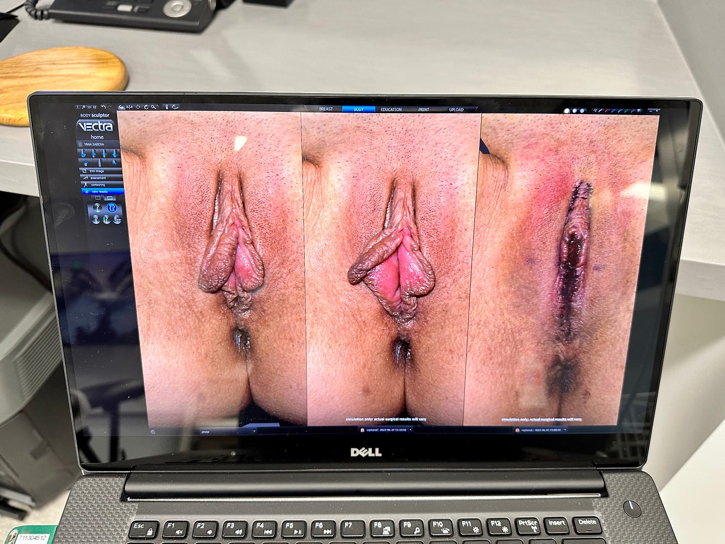
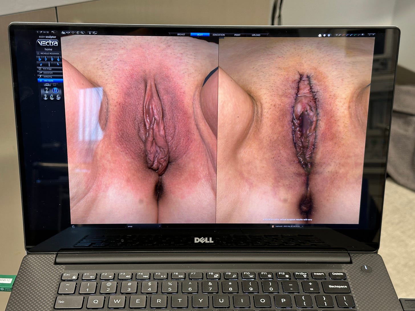
I have collected the world’s first set of 3D photos illustrating the results of my labial and vaginal surgeries and majora augmentation. They show the appearance before surgery, often immediately after surgery, and appearance approximately 6-8 weeks after surgery. A great deal of time and effort has been exerted by my patients and staff to obtain this landmark collection. Thank you to Canfield Scientific for their Vectra 3D system that made this all possible. This is truly amazing. You can view the changes over time at just about all the usable angles. I use this in my practice to evaluate my results from what the patient sees and her point of view. We teach this in our Alinsod Fellowship programs.
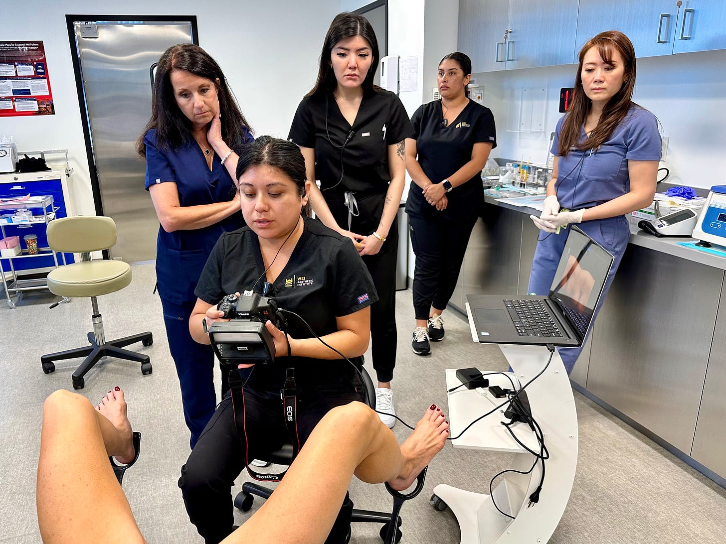
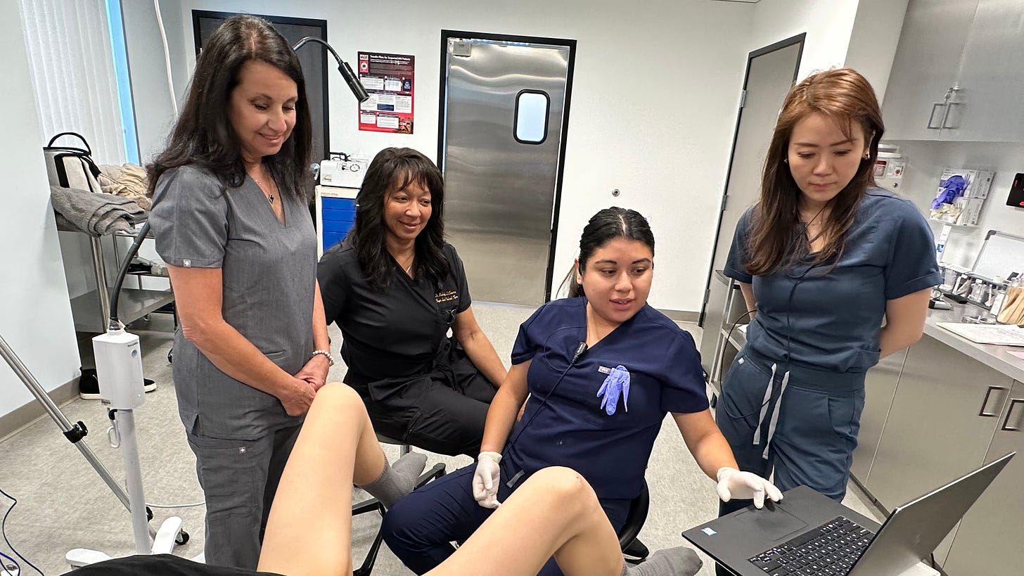
Marisol shows how simple 3D photography is done with a special camera lens system able to take pictures and blend them into a rotatable set.
The computer then stores the photos and allows comparison over time at all the angles of rotation.
Here is a set of Before and Immediately After photos.
Imagine your Before and After photos side by side and rotating them to the view you want so see.
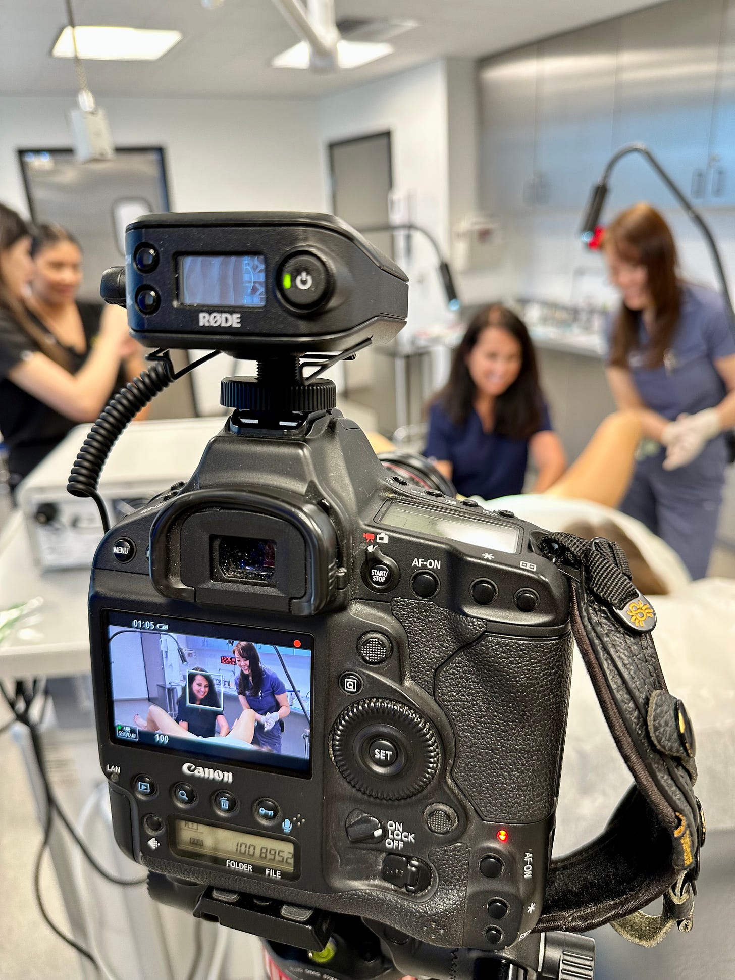
We videotaped our surgeries and procures for more future Gynflix content.
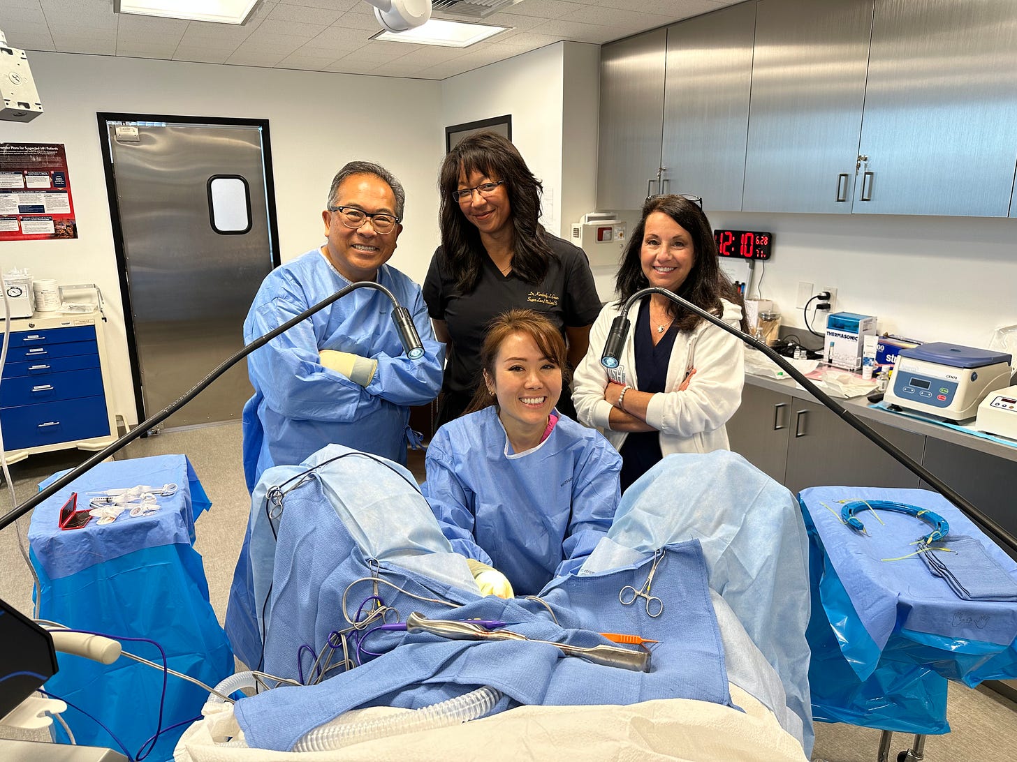
Dr. Wei and I just finished our successful and fun training week with new Fellows Kimberly Evans from Houston and Francine Fradella from Long Island.

Click Here for more: 3-D Labial Photography Cases as seen on www.urogyn.org. Join www.gynflix.com for the best photos and videos available for learning.

Red Alinsod, MD (www.urogyn.org and www.gynflix.com)

Cosmetic Gynecology Newsletter is a reader-supported publication. To receive new posts and support my work, become a paid subscriber. It is only ten buck a month.

Leave a comment

Share

Cosmetic Gynecology Newsletter
Authors
Cosmetic Gynecology Newsletter